Emilia Aram , David C. Flanigan, MD, Nathan J. Castro, PhD, Doug Snell, Luke Aram
Purpose: Osteochondral defects can present a significant source of pain and impairment of a patient’s quality of life and current solutions are inadequate:
- Microfracture is known to have inconsistent results and lack durability
- Matrix-Induced Autologous Chondrocyte Implantation and biological solutions are expensive and have a long recovery time
In this study, we evaluated the ease of implantation and fixation strength of three fixation designs integrated within Nanochon’s Chondrograft cost-effective cartilage repair implant for shortened recovery time.
Materials and Methods: Three fixation designs were evaluated on human and porcine cadaver knees (Figure 2). Surgeons rated implant designs and tooling from one (good) to five (bad) based on ergonomics, appropriate size, ease of use, accuracy, and repeatability. Fixation strength was tested by applying sutures to the implant and using a force gauge to pull out the implant.
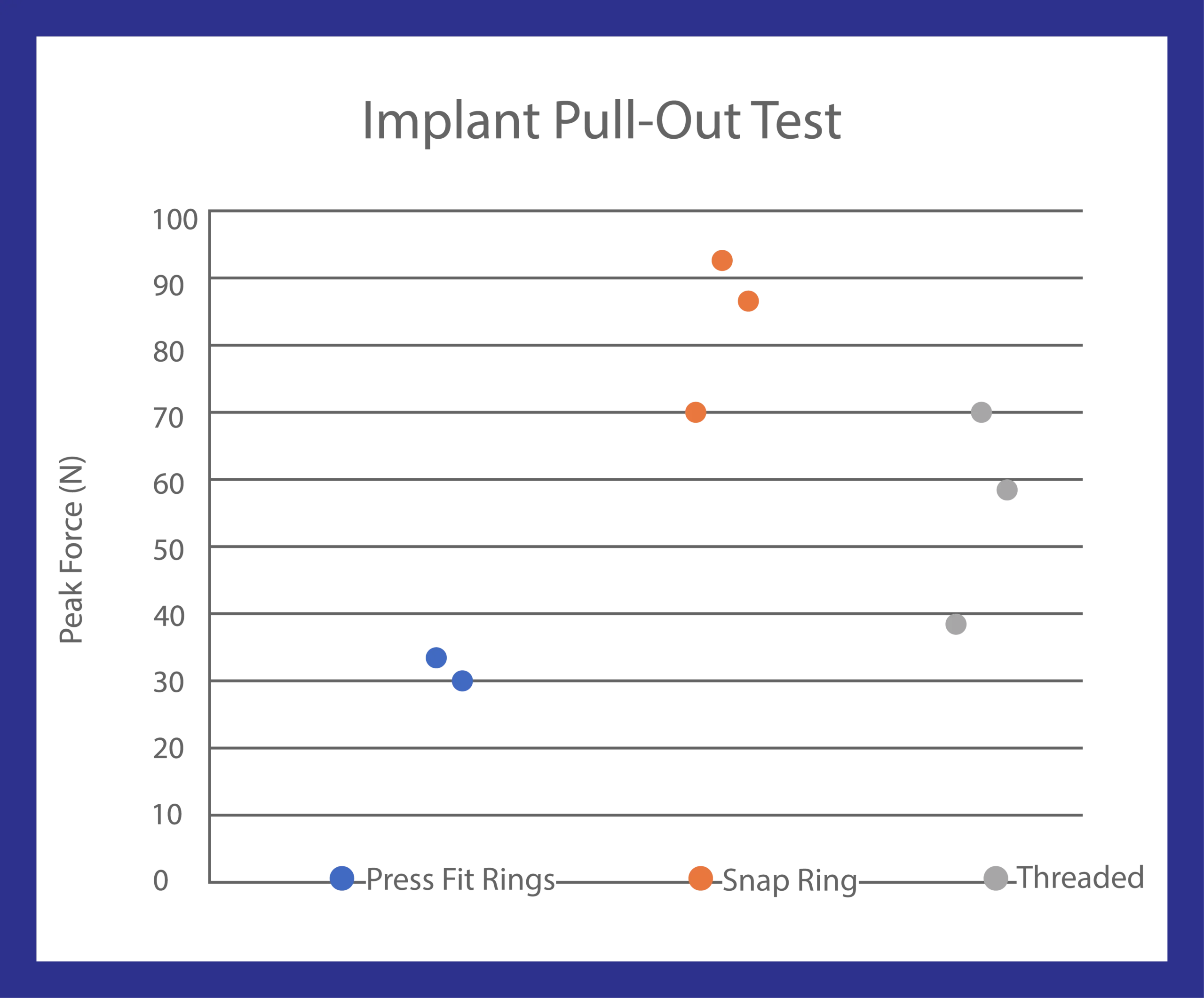
Results: The Press Fit Ring was easiest to use (1.6) and most accurate (1.2) while allowing for single-hand use when compared to Snap Ring (1.0 and 1.7, respectively). Additionally, the Press Fit Ring was found to be more ergonomic than Snap Ring (1.2 and 1.4 respectively), as well as slightly more repeatable (Press Fit Ring 1.2 and Snap Ring 1.3). Snap Ring scored lowest (best) for instrumentation size followed by Press Fit Ring (2.4 and 3.0 respectively) (Figure 3). Snap Ring also had the highest average pull-out force, 83.4 N, compared to 55.7 N for the threaded fixation and 29.2 N for the Press Fit Ring (Figure 4).
Conclusions: Surgeons unanimously preferred Press Fit Ring from the overall ease of use, and, as a result of this study, will continue to be evaluated in animal studies. Regarding pull-out force, all three implant designs met the acceptance criteria (>23.7 N). Large animal testing is planned to demonstrate Press Fit Ring implant’s durability.
Figure 1. Depiction of types of chondral defects.
Chondral defect, partial thickness

Chondral defect, full thickness

Osteochondral
defect

Figure 2. CAD renderings of the three fixation designs. A) Snap Ring: Includes an integrated ring that can be compressed, inserted into a prepared socket in the knee joint, and released such that the ring interlocks with the socket. B) Press Fit Ring: Includes two rings that can be press-fit into a prepared socket. C) Threaded: can be rotated into place like a screw.
A) Snap Ring

B) Press Fit Ring
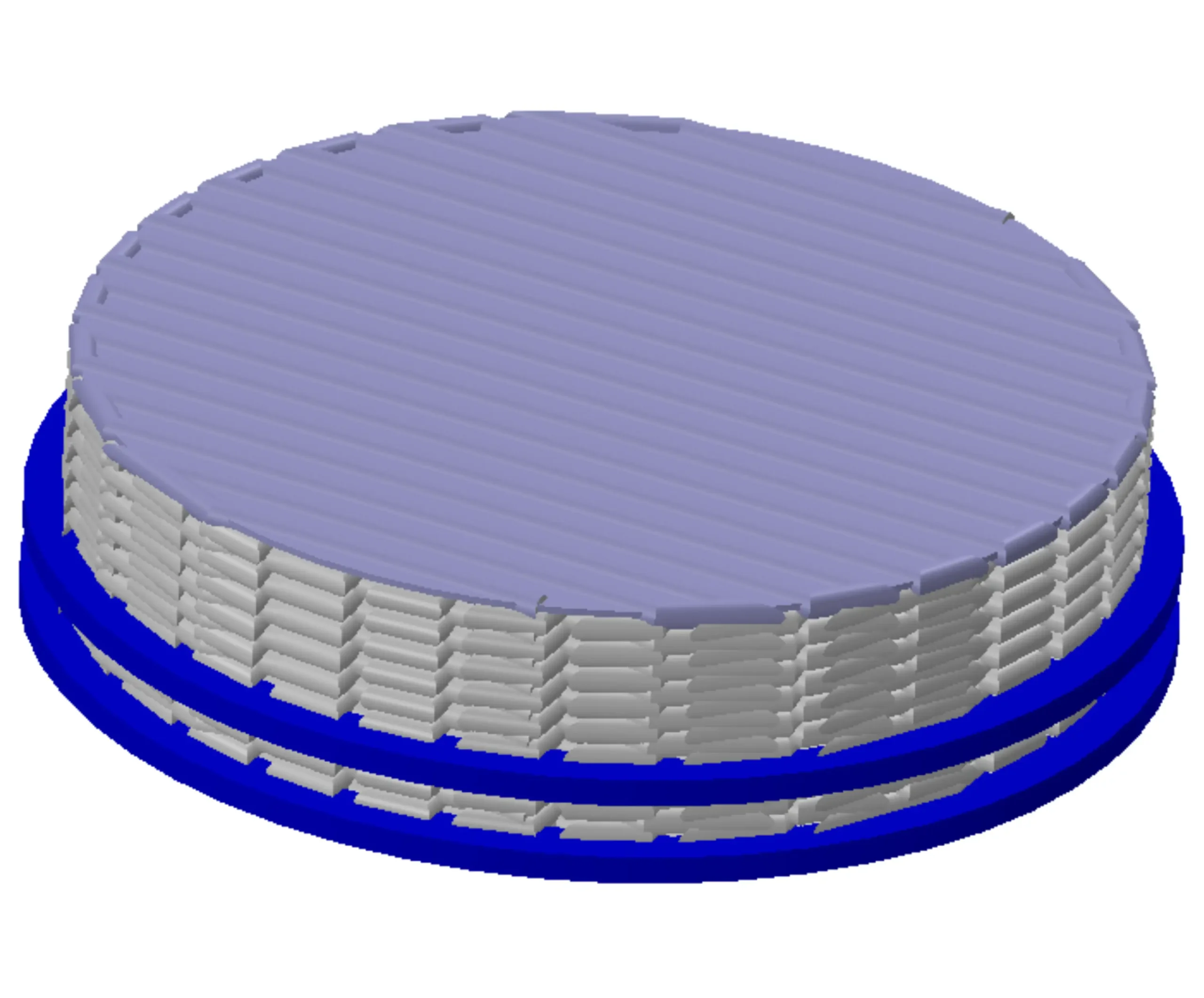
C) Threaded

Figure 3. Actual fixation devices evaluated on human and porcine cadaver knees. A) Snap Ring was found to have the best instrumentation size and had the highest average pull-out force. B) Press Fit Ring was found to be easiest to use, most accurate, more ergonomic and slightly more repeatable C) Threaded fixation device
A) Snap Ring
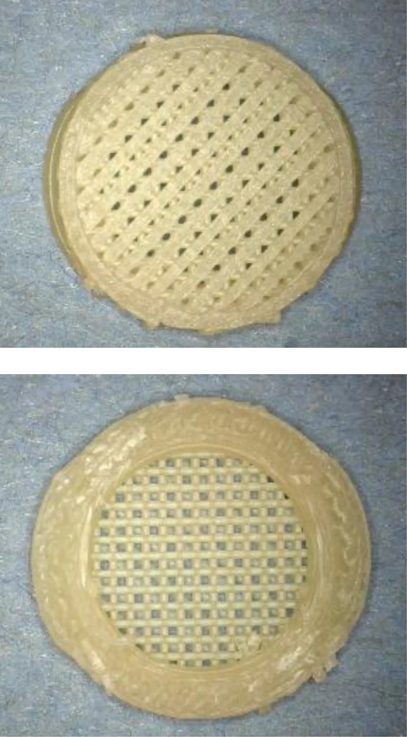
B) Press Fit Ring
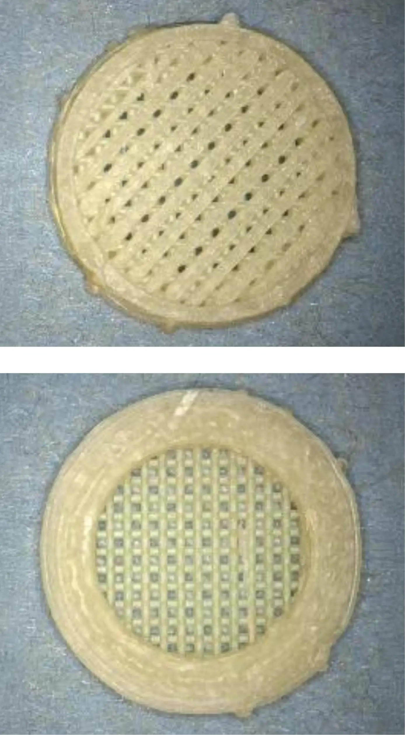
C) Threaded

Figure 4. Graphical representation of results of implant pull-out test.
Presented at 18th ICRS World Congress “Joint Revolution”